
Introduction - case 1407 (ultrasonographic picture 5)
|
|

|
|
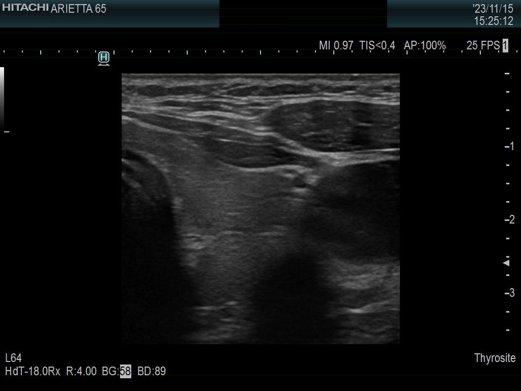
Left lobe, transverse scan. This lobe is also minimally hypoechoic. A connective tissue runs through the lobe which makes the appearance of the dorsal part nodular.