
Introduction - case 1407 (ultrasonographic picture 11)
|
|

|
|
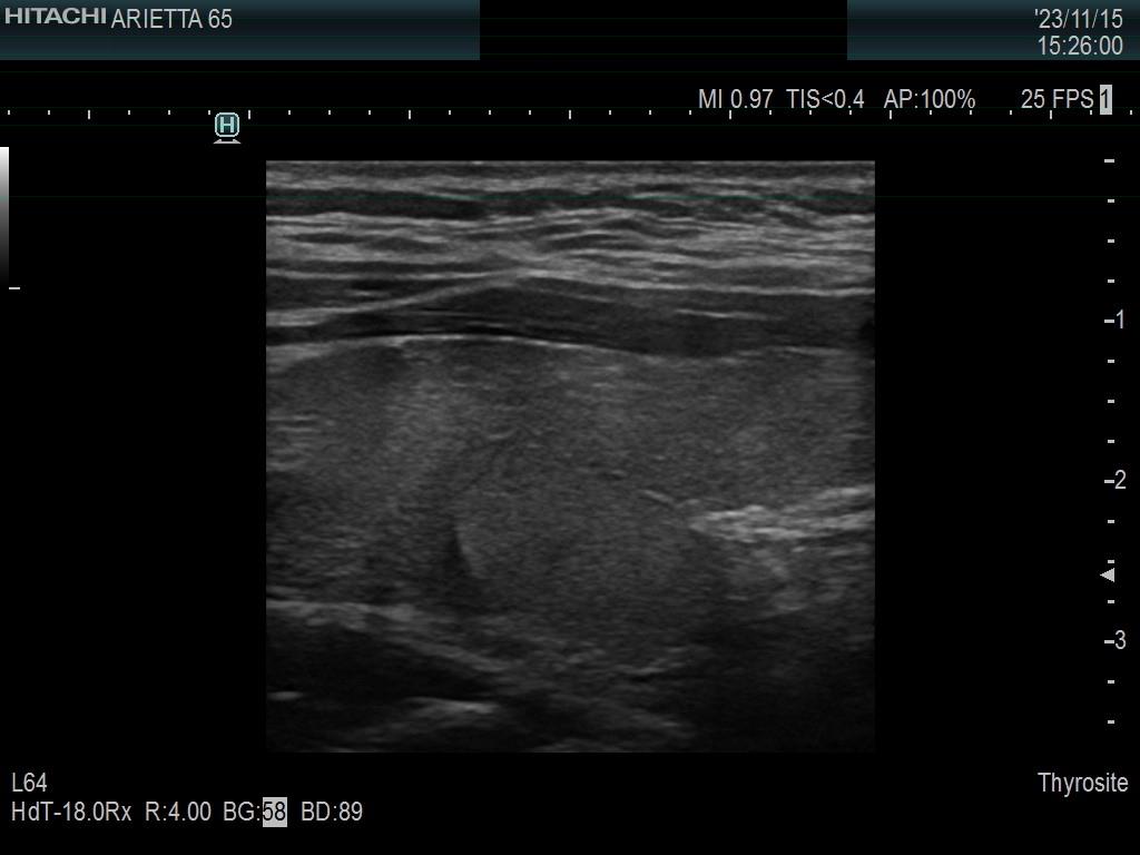
Left lobe, longitudinal scan. Two circumstances explain that at first sight we can judge the dorsal part of the lobe as a nodule. The fragmented connective tissue running ventral and the hypoechoic areas upper and lower (i.e. left and right in the image) to this part of the lobe.