
Introduction - case 162 (ultrasonographic picture 3)
|
|

|
|
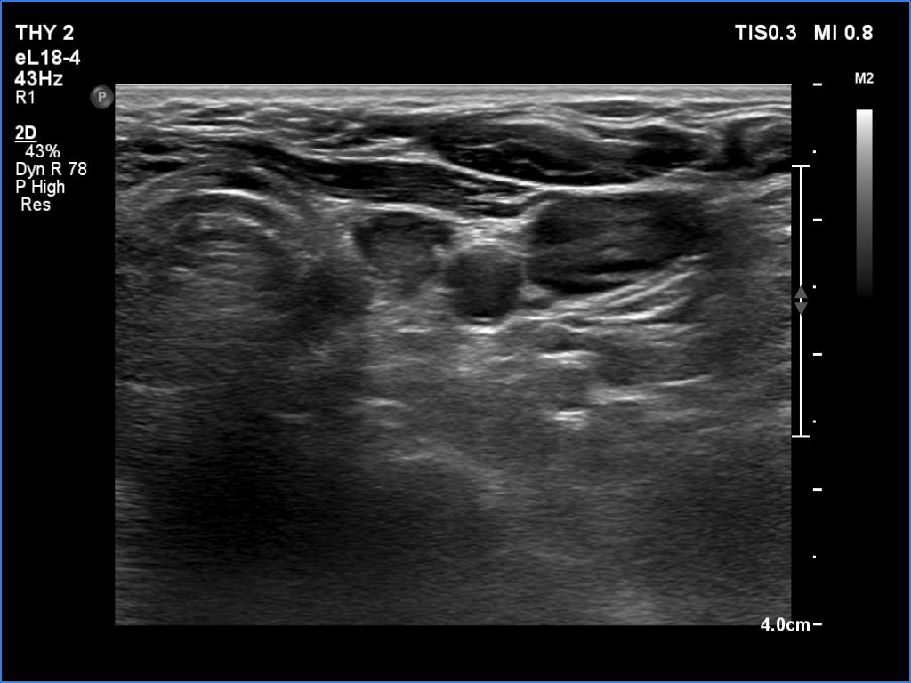
Left lobe, transverse view - with less harmonic settings. There is a hypoechoic mass in the left throid bed. On this view, this could be even a normal finding.