
Introduction - case 487 (ultrasonographic picture 1)

|
|
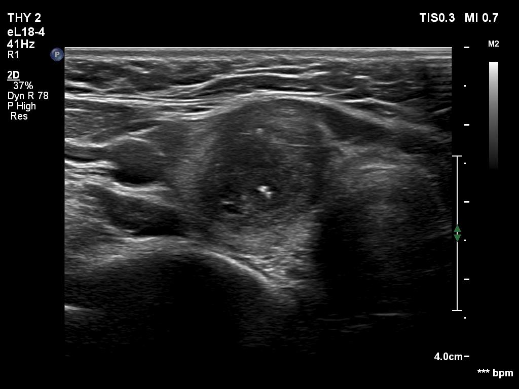
Right lobe, transverse view. There is a moderately hypoechoic nodule which has very hypoechoic part and a few cystic areas, as well. Note taller-than-wide shape.