
|
|
Introduction - case 745
|
|


Clinical presentation: A 32-yr-old woman was referred for evaluation of a thyroid nodule which was discovered by the patient herself. She noticed a lump in the middle of the neck a few months ago.
Palpation: a hard nodule in the isthmus and another firm lesion in the right lobe.
Laboratory tests: TSH 1.89 mIU/L.
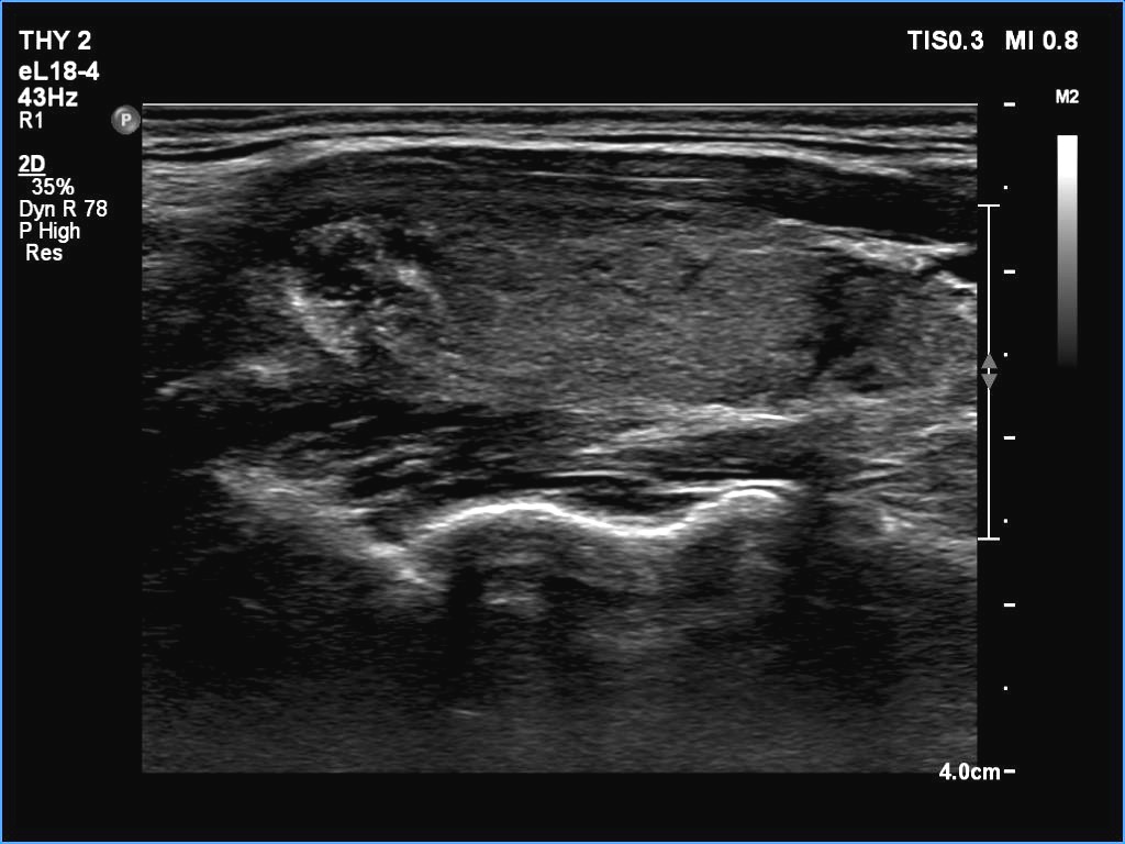
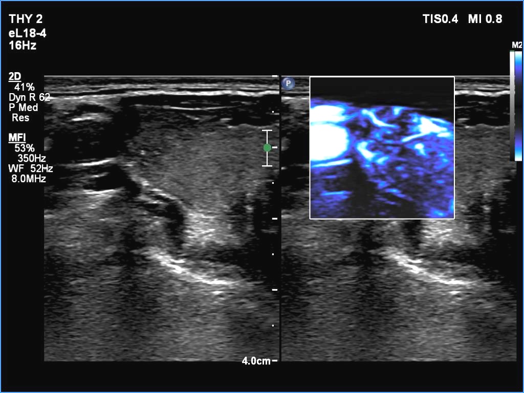
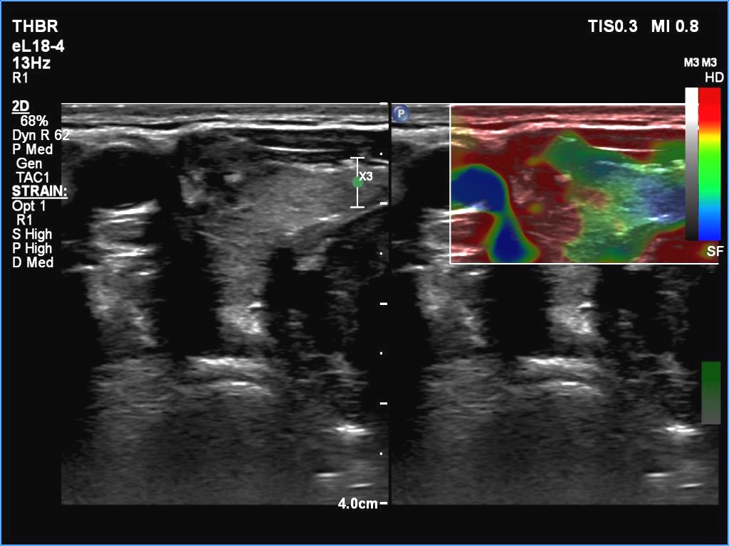
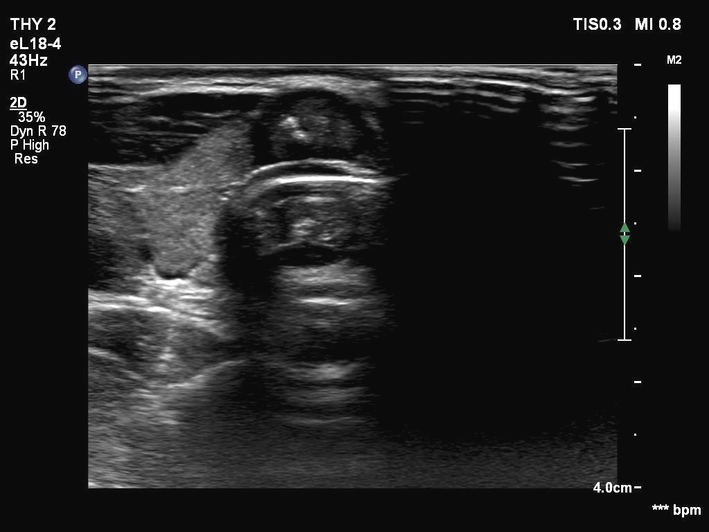
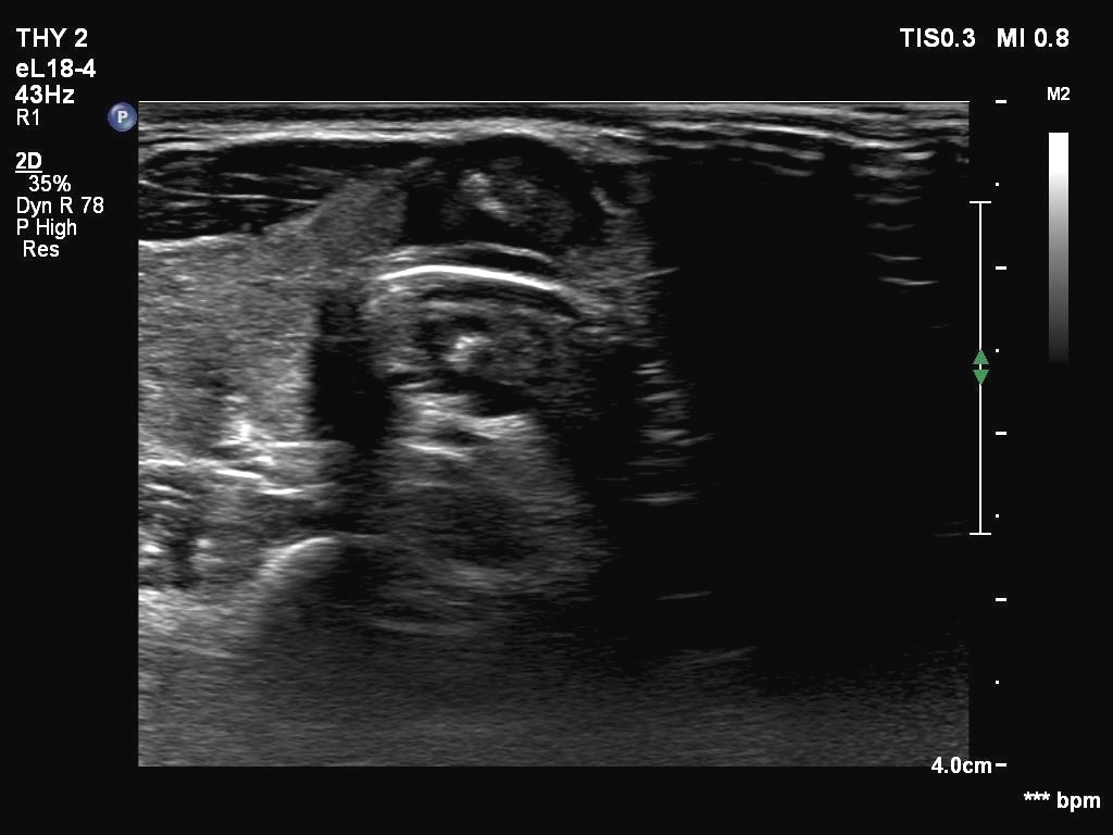
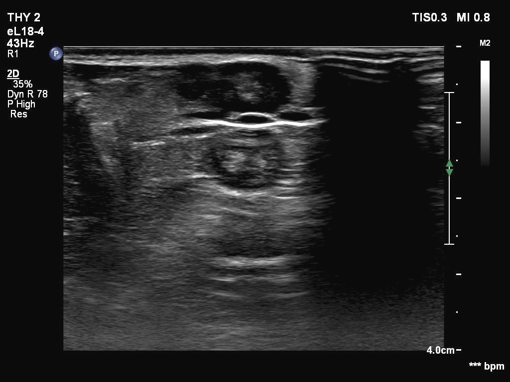
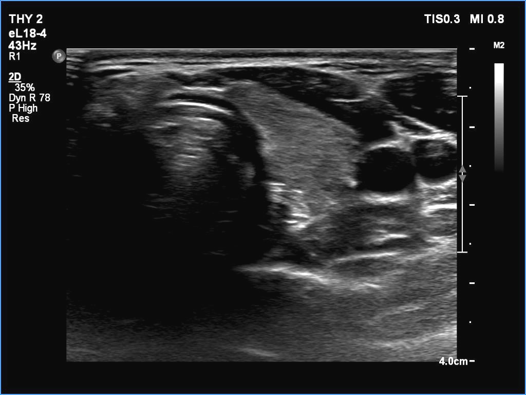
Ultrasonography. The thyroid was echonormal. There were two partly deeply hypoechoic nodules, one in the ventrolateral part of the right lobe and another one in the isthmus. Both had irregular margins and punctate echogenic foci. There was a mirror image artifact of the lesion in the isthmus.
FNA of the nodule in the right lobe resulted in papillary cancer while cytology of the isthmis lesion did in suspicion of papillary cancer.
Histopathology resulted in T2b multifocal papillary cancer.
Comments.
-
Both nodules had multiple suspicious features: beside deep hypoechogenicity and irregular borders, microcalcifications should be also considered. The nodule in the right lobe presented also three possible sonographic signs of a possible extrathyroidal spread: the pseudocapsule of the thyroid was not intact, the lesion had both abutting and butting contours.
-
According to the literature, the mirror image artifact is not an exceptional finding. Nevertheless, such a reflection very similar to the original is quite rare. The prerequisite of this phenomenon is the flattening of the trachea' wall as is observed in this case. The explanation is as follows: 'The primary beam reflects from such a surface (e.g. diaphragm) but instead of directly being received by the transducer, it encounters another structure (e.g. a nodular lesion) in its path and is reflected back to the highly reflective surface (e.g. diaphragm). It then again reflects back towards the transducer.' (Source: radiopedia.org.)