The borders of the nodule - case conp 040 (ultrasonographic picture 6)
|
|

|
|
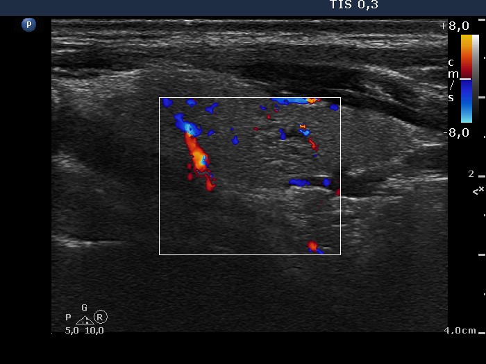
Left lobe, longitudinal view, color Doppler mode. The lesion presents signs of perinodular and intranodular blood flow.

Papillon Course
2024-25
Nodule' borders
|
|
|
|
Left lobe, longitudinal view, color Doppler mode. The lesion presents signs of perinodular and intranodular blood flow.