
The composition of the nodule - case 399 (ultrasonographic picture 6)
|
|

|
|
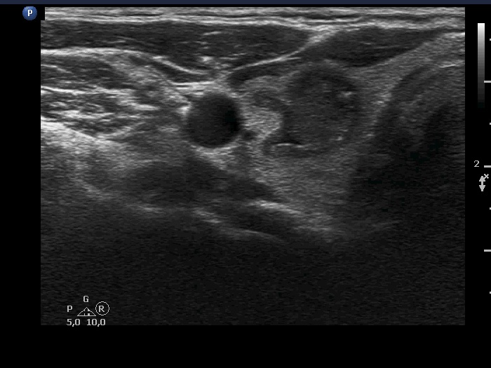
Right lobe, transverse scan - after the removal of 2.5 mL cystic content. The nodule borders became lobulated. The intranodular echogenic figures are mostly dorsal to tiny cystic areas.