
Intranodular hyperechogenic figures - case 386
30 month after the initial examination (ultrasonographic picture 1)

|

|

|
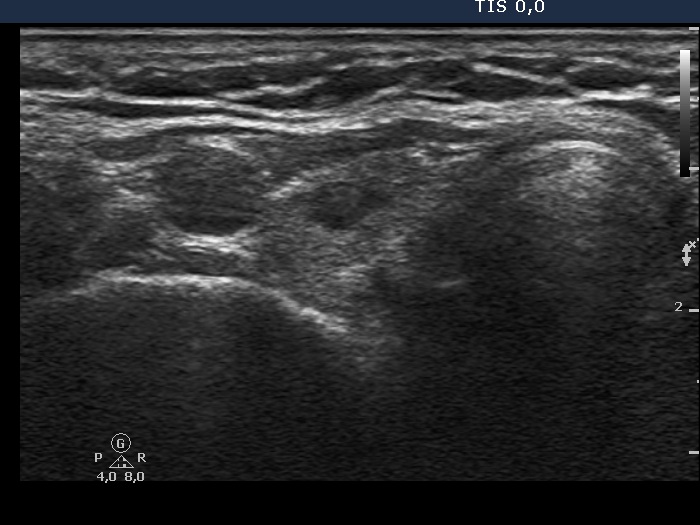
Right lobe, transverse scan. There is a small hypoechogenic lesion in the right lobe.


