
|
|
The echogenicity of the nodule - case 1177
|
|

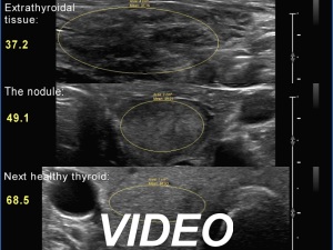
Clinical presentation: A 57-year-old woman was referred for aspiration cytology. Hypothyroidism was revealed by a routine blood test for 6 weeks. Thereafter, a hyperechoic nodule was found on ultrasound.
Palpation: Both lobes were firm. No obvious nodule was palpated.
Laboratory tests: TSH 2.39 mIU/L on daily 75 microgram levothyroxine.
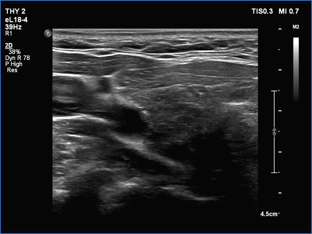
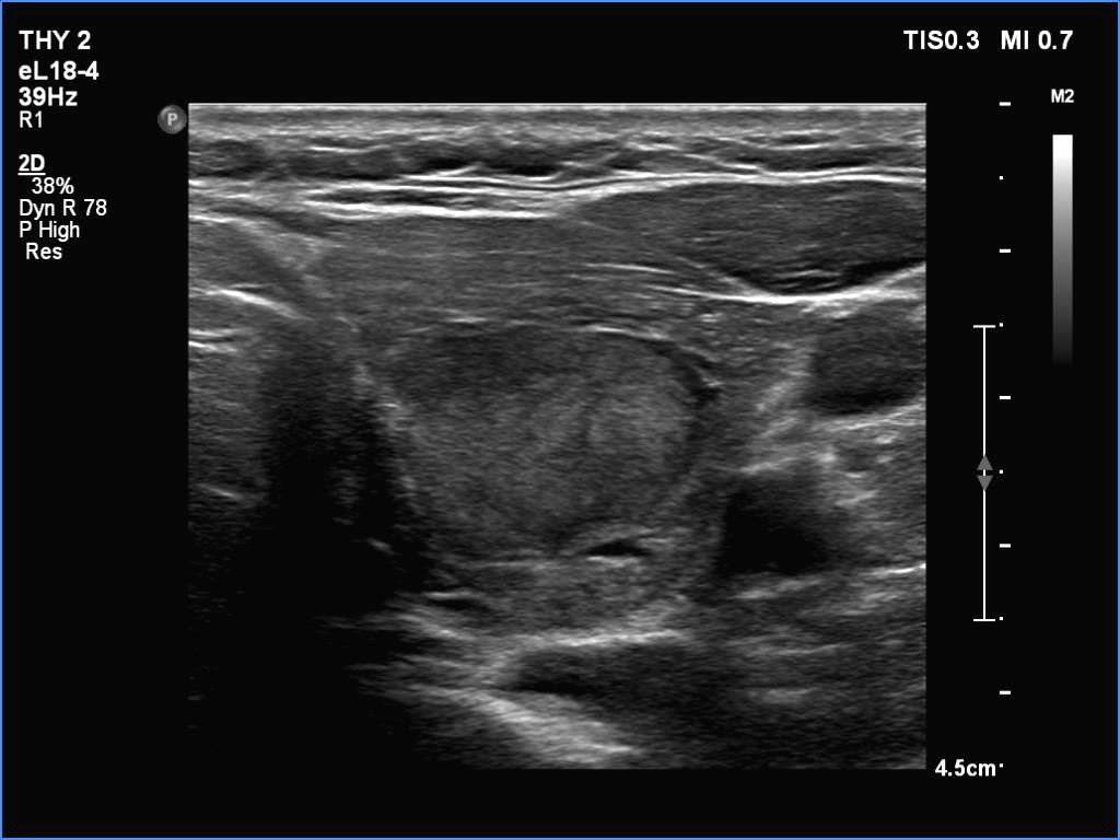
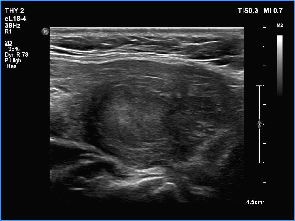
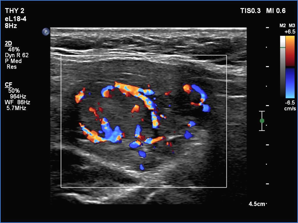
Ultrasonography. The thyroid was partly minimally/moderately, partly very hypoechoic. There was a nodule in the middle of the left lobe. It was lighter than the extranodular part but darker than a normal, healthy thyroid.
FNA resulted in benign lesion.
Comments. All but one society suggest to compare the nodule' echogenicity to the non-nodular part. In this case, the nodule should be regarded as iso/hyperechoic. If we accept the suggestion of the 2023 ETA guideline, then we should consider the nodule as minimally/moderately hypoechoic because the reference tissue in the ETA is the normal, healthy thyroid and not the diseased, extranodular part.