
The echogenicity of the nodule - case 2118 (ultrasonographic picture 4)
|
|

|
|
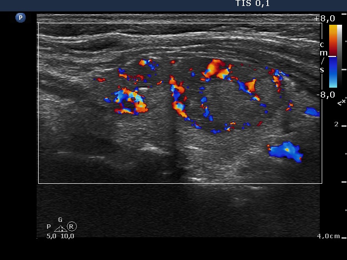
Right lobe, longitudinal scan, color Doppler mode. The upper nodule (left in the image) shows intranodular vascularization while the larger, lower nodule (right in the image) presents both perinodular and intranodular vascularization.