
The shape of the nodule - case 2052 (ultrasonographic picture 1)

|
|
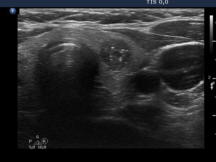
Left lobe, transverse scan. There is a hypoechoic nodule which shows taller-than-wide shape. In this image, the intranodular echogenic figures seem to be microcalcifications.