
Study on 100 consecutive patients with thyroid nodule - case 023
One year before the present examination (ultrasonographic picture 3)
|
|

|
|
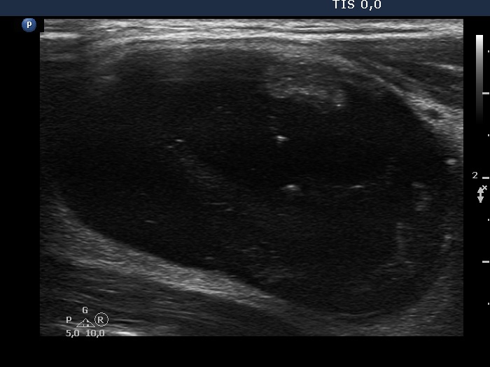
Right lobe, longitudinal scan. Note an isoechoic solid part in the ventral wall.