
Study on 100 consecutive patients with thyroid nodule - case 030 (ultrasonographic picture 12)
|
|

|
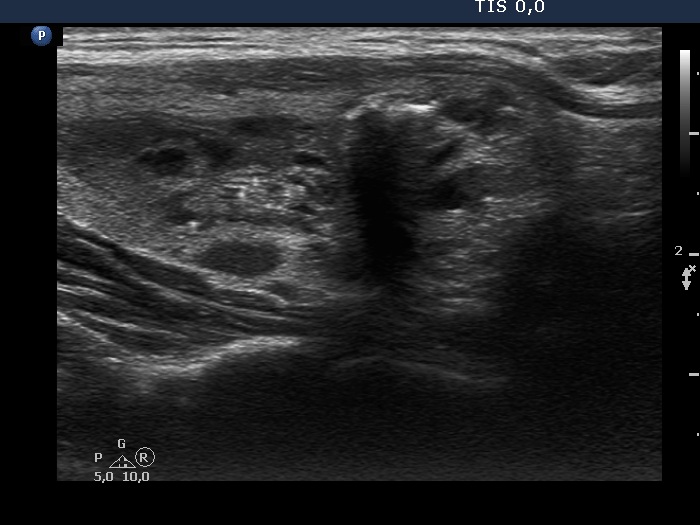
Right lobe, another longitudinal scan - after aspirating 6 mL brown fluid. On this view, the echogenic granules are more likely back wall figures.