
Study on 100 consecutive patients with thyroid nodule - case 035 (ultrasonographic picture 3)

|

|

|
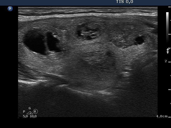
Right lobe, longitudinal scan. The bright hyperechogenic intranodular figures correspond partly to comet-tail artifact partly to back wall figures or fibrosis.


