
Study on 100 consecutive patients with thyroid nodule - case 037 (ultrasonographic picture 2b)

|

|

|
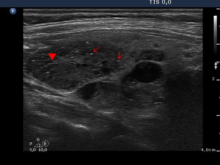
Right lobe, longitudinal scan. The bright hyperechogenic granules (arrows) seem to be at first sight microcalcifications. However, the presence of a few hyperechogenic lines (arrowheads) challenges this view: these figures might be presentations of connective tissue or back wall cystic figures.


