
|
|
Study on 100 consecutive patients with thyroid nodule - case 052
|
|


Clinical presentation: A 43-year-old woman was referred for evaluation of a nodular goiter detected on PET CT scan which was performed on staging of a colon cancer.
Palpation: The left lobe had a not firm nodule.
Hormonal evaluation indicated euthyroidism (TSH 1.93 mIU/L).
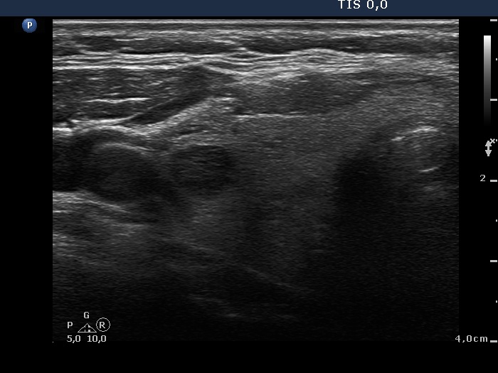
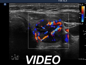
Ultrasonography. The thyroid was echonormal. There were several small discrete partly echonormal, partly moderately hypoechogenic-cystic lesions in the right lobe. The left lobe presented a large nodule composed of peripheral echonormal solid part and a central complex moderately hypoechogenic-cystic area. The hypoechogenic part had a partly chaotic, increased vascular pattern.
Cytology of resulted in benign cystic lesion.
Suggestion: repeat ultrasound in a year.