
|
|
Study on 100 consecutive patients with thyroid nodule - case 067
|
|


Clinical presentation: A 65-year-old woman came to a follow-up. We met the patient first, 17 years ago., At this time, a benign multinodular goiter was diagnosed. A nodule with the dimensions of 18x13x45 mm (5.51 mL in volume) was found in the left lobe which dimensions were 25x15x50 mm (9.81 mL). The patient has regularly checked her thyroid. She had no complaints.
Palpation: a moderately firm nodule in the left lobe.
Functional state: euthyroidism (TSH 0.58 mIU/L).
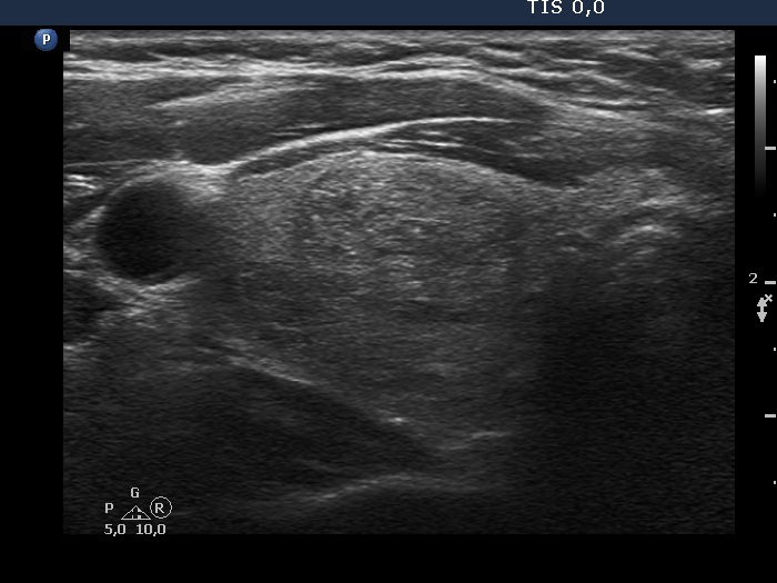
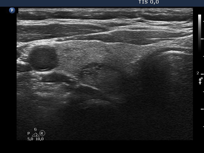
Ultrasonography. The thyroid was echonormal and had multiple nodules, mostly minimally hypoechoic or echonormal. The dimensions of the left lobe were 27x15x57 mm (12.1 mL in volume). This lobe had a minimally hypoechoic-echonormal nodule which presented halo sign. The dimensions of the nodule were 22x13x53 mm (7.93 mL in volume). Compared with the first examination for 17 years, the volume of the nodule increased by 23% while the nodule did by 43.9%. (The height of the patient was 168 cm, the weight was 86 kg.)
Aspiration cytology performed from the large nodule resulted in benign colloid goiter.
Suggestion: ultrasound in three years.
Comment. There are several important lessons to be learned from this case study.
-
The more important is that not the size of the nodule but the size of the lobe which influences our decision when indicating surgery in the case of thyroid enlargement. The volume of the lobe should be compared to the BMI. The enlargement of the lobe in this case should be regarded only minimal. It means that surgery is not indicated.
-
Great proportion of nodules increase in size. We should never forget, that initially all nodules had a maximal diameter of 1 mm. Be aware of the following: if a lesion grows from 1 mm to 10 mm, it means a 1000-fold increase in volume. a further growing to 20 mm means an 8-times increase in volume.