
Consecutively operated patients with autoimmune thyroid disease - case 6 (1424) (ultrasonographic picture 2)
|
|

|
|
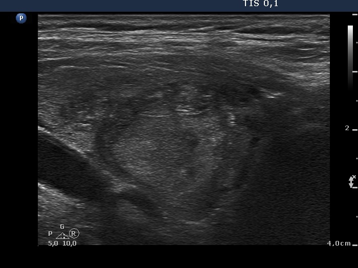
Right lobe, longitudinal view. The small moderately hypoechogenic area presenting echogenic figures might have some oncological significance. This lesion has both echogenic lines and granules, therefore these figures correspond to connective tissue.