
Discrete lesion or nodule in Hashimoto's thyroiditis - case 16 (797) (ultrasonographic picture 3)
|
|

|
|
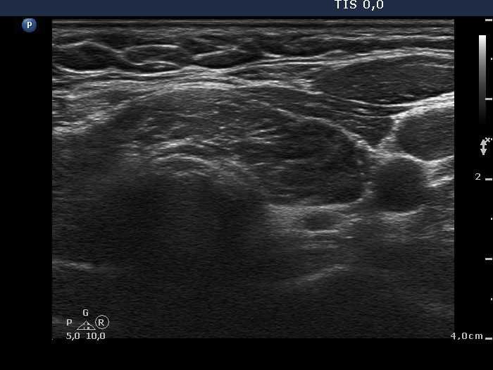
Isthmus and lower part of the left lobe, transverse view. Note the synchronous presence of echogenic granules and lines; this pattern corresponds to connective tissue.