
100 consecutive cases of papillary cancer - case 002 (ultrasonographic picture 2)
|
|

|
|
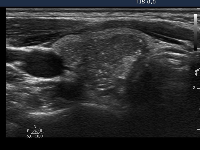
Right lobe, transverse scan, enlargement. Part of the hyperechogenic figures might correspond to microcalcification.