
100 consecutive cases of papillary cancer - case 016 (ultrasonographic picture 9)
|
|

|
|
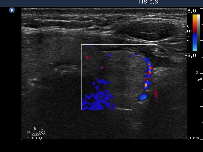
Left lobe, transverse scan, color Doppler mode. The nodule present perinodular blood flow. The irregular blue patches at the left lower corner of the insert are technical artifacts.