100 consecutive cases of papillary cancer - case 027 (cytologic picture 18)
|
|

|
 |
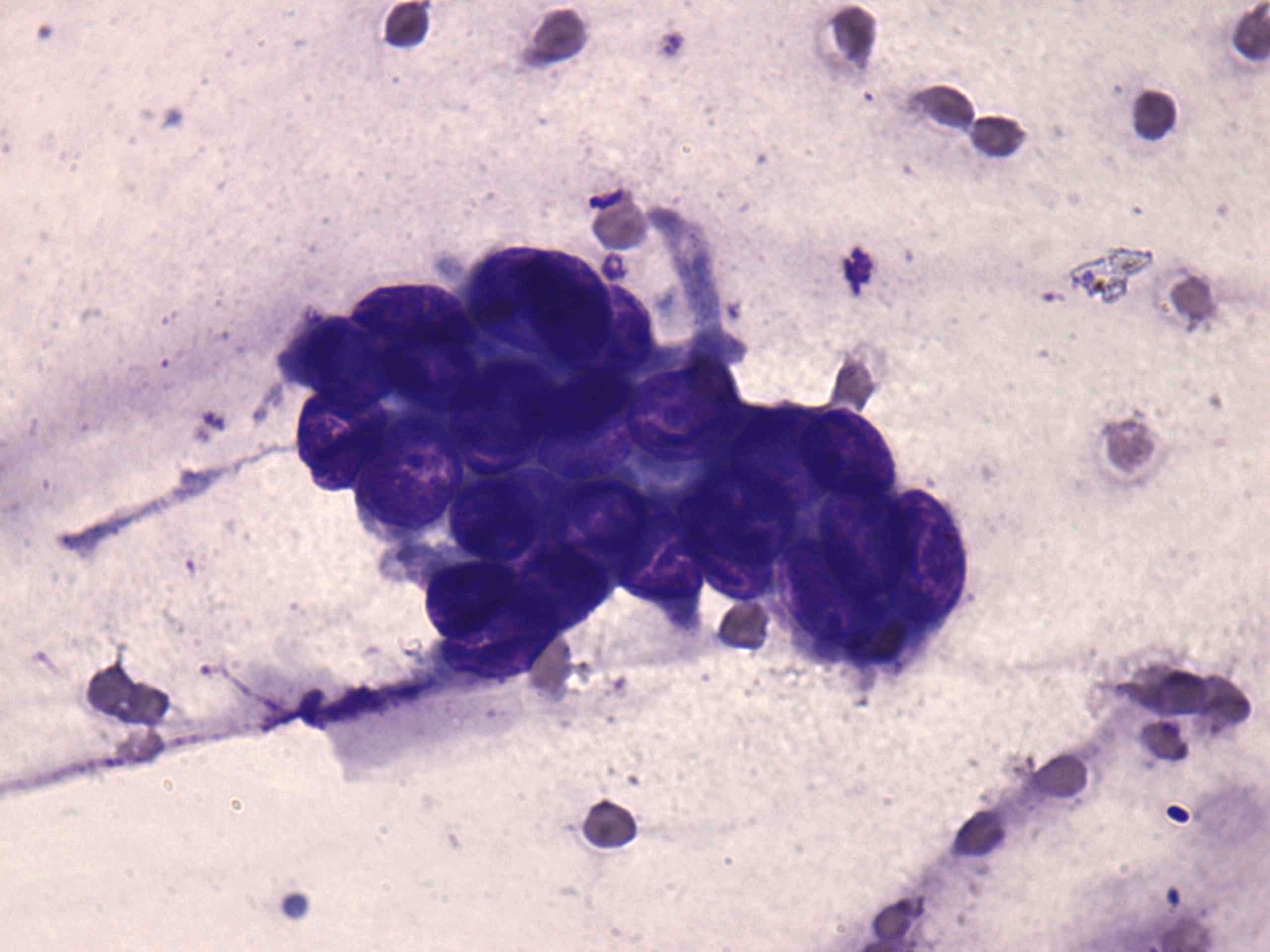
Wright-Giemsa staining, 1000x. A tip of a papillary fragment can be seen in the image.

Papillon Course
2024-25
Supplementum
|
|
|
|
Wright-Giemsa staining, 1000x. A tip of a papillary fragment can be seen in the image.