
100 consecutive cases of papillary cancer - case 040 (ultrasonographic picture 6)
|
|

|
|
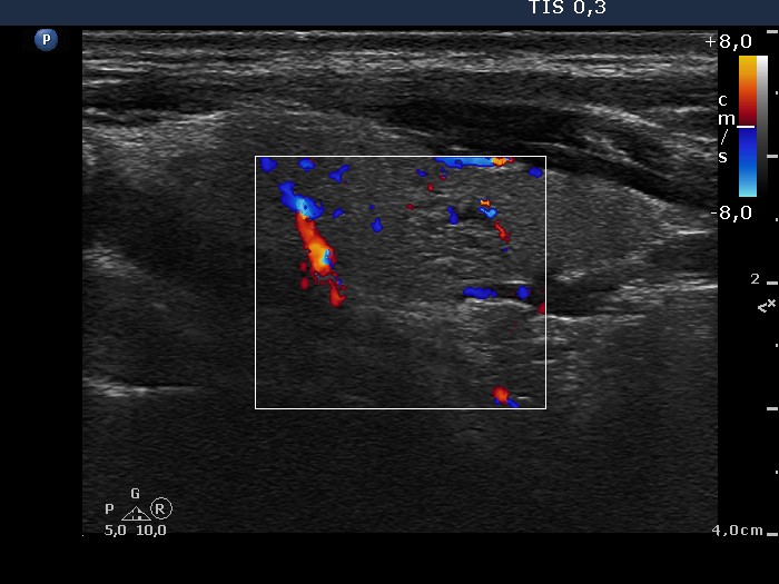
Left lobe, longitudinal view, color Doppler mode. The lesion presents signs of perinodular and intranodular blood flow.