
100 consecutive cases of papillary cancer - case 080 (ultrasonographic picture 3)
|
|

|
|
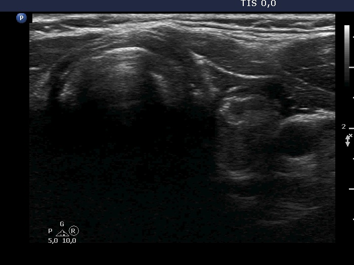
Upper part of the left lobe, transverse view. There is a dominantly isoechoic, heterogeneous nodule in the ventral part of the lobe.