100 consecutive cases of papillary cancer - case 089 (ultrasonographic picture 5)
Follow-up examination a year later
|
|

|
|
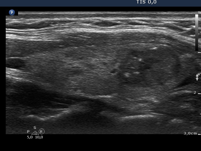
Left lobe, longitudinal scan, enlargement. Note the irregular borders.

Papillon Course
2024-25
Supplementum
Follow-up examination a year later
|
|
|
|
Left lobe, longitudinal scan, enlargement. Note the irregular borders.