100 consecutive cases of papillary cancer - case 096 (ultrasonographic picture 8)
|
|

|
|
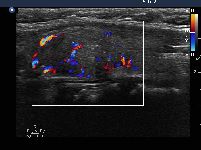
Left lobe, longitudinal scan, color Doppler mode. The lesion has both perinodular and intranodular vascularity.

Papillon Course
2024-25
Supplementum
|
|
|
|
Left lobe, longitudinal scan, color Doppler mode. The lesion has both perinodular and intranodular vascularity.