
|
|
100 consecutive cases of papillary cancer - case 098
|
|


Clinical presentation: A 74-year-old woman was referred for aspiration cytology of a thyroid nodule which was discovered by the patient herself six months ago. She noticed that the mass has increased in size.
Palpation: a firm nodule in the right lobe.
Laboratory test: TSH 3.98 mIU/L.
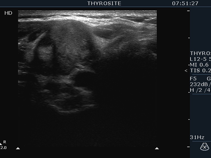
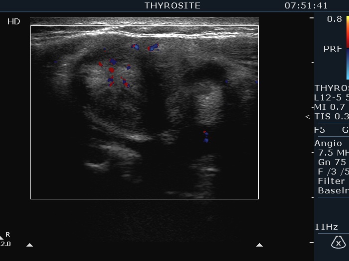
Ultrasonography. The thyroid was echonormal and had multiple nodules. The largest lesion in the right lobe was minimally hypoechoic had irregular borders and presented all three possible signs of an extrathyroidal spread. The vascularity was scanty. The largest nodule in the left lobe was also remarkable due to numerous echogenic lines and granules which were related to ventral cystic areas. So, these were back wall figures.
Cytology resulted in papillary cancer.
Total thyroidectomy was performed. Histopathology disclosed a T4N1b papillary cancer according to the nodule in the right lobe. The tumor invaded the soft tissue ventral to the thyroid. The largest nodule in the left lobe proved to be follicular adenoma.