
100 consecutive cases of papillary cancer - case 099 (ultrasonographic picture 7)
|
|

|
|
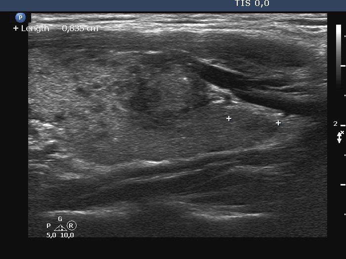
Left lobe, longitudinal scan. Due to technical reasons, the nodule is darker in longitudinal scan. Note a microcalcification.