
|
|
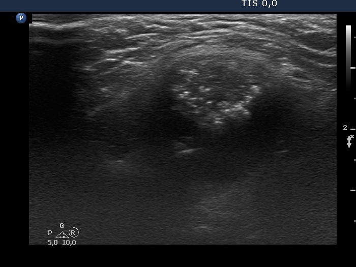
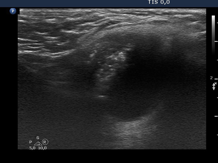
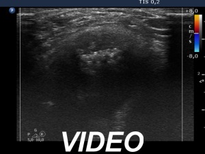
The composition of the nodule - case 51
|
|


Clinical presentation: A 33-year-old man requested a second opinion. He has noticed a lump in the neck for a year. Aspiration cytology resulted in benign lesion.
Palpation: a moderately firm mass in the middle of the neck above the thyroid.
Result of blood test: TSH 0.64 mIU/L.
Ultrasonography. The thyroid was echonormal and had a minimally-hypoechogenic lesion in the upper-ventral part of the right lobe. It was equivocal whether this area was composed of two nodules or was in fact one nodule with lobulated margins. According to the mass in the middle part of the neck, a cystic lesion was found. It had a hypoechogenic solid part presenting numerous punctate echogenic foci, great proportion of them were related to tiny ventral cystic areas.
Aspiration cytology was performed both form the neck mass and the thyroid nodule. The former resulted in a very cellular smear without pathognomonic nuclear signs of papillary carcinoma.
We performed wash-out thyroglobulin determination which yielded > 486 microgram/L. Our combined clinical-ultrasound-cytological diagnosis was suspicion of papillary thyroid carcinoma. The cytology of the thyroid nodule in the right lobe proved to be benign.
Histopathology not yet known.