
|
|
Intranodular hyperechogenic figures - case 1108
|
|


Clinical presentation: A 34-year-old woman was referred for follow-up examination on 8th gestational week. She was known to have hypothyroidism for 2 years which was replaced by 50 microgram levothyroxine. Thyroid autoantibody levels were formerly elevated: aTPO 177 U/mL, anti-hTg 278 U/mL.
Palpation: no abnormality.
Hormonal investigation indicated euthyroidism with lower TSH-level 0.16 mIU/L, FT4 14.2 pM/L.
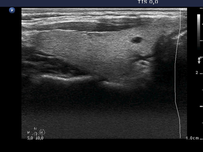
Ultrasonography: The thyroid was echonormal. There were numerous hypoechoic, well-circumscribed, small lesions, some of them presented comet-tail artifact. The left lobe contained less hypoechogenic and less well-demarcated areas, as well. The former corresponded to insignificant lesions while the latter were signs of the underlying autoimmune thyroiditis.
Suggestion: we did not change the replacement dose because in the first trimester the TSH may decrease normally to 0.1 mIU/L. The THS was in the normal range later in the course of pregnancy, 2.01 mIU/L at 21st and 2.28 mIU/L at 33rd week of gestation.