
|
|
Intranodular hyperechogenic figures - case 1139
|
|


Clinical presentation: A 55-year-old woman was referred for aspiration cytology. She has a right lobectomy 10 years ago. At that time, a relatively small cystic nodule was already in the left lobe which was not operated. She noticed a lump in the left thyroid several weeks before the present examination.
Palpation: an elastic nodule in the left lobe.
Functional state: euthyroidism (TSH 1.7 mIU/L, aTPO 641 U/mL).
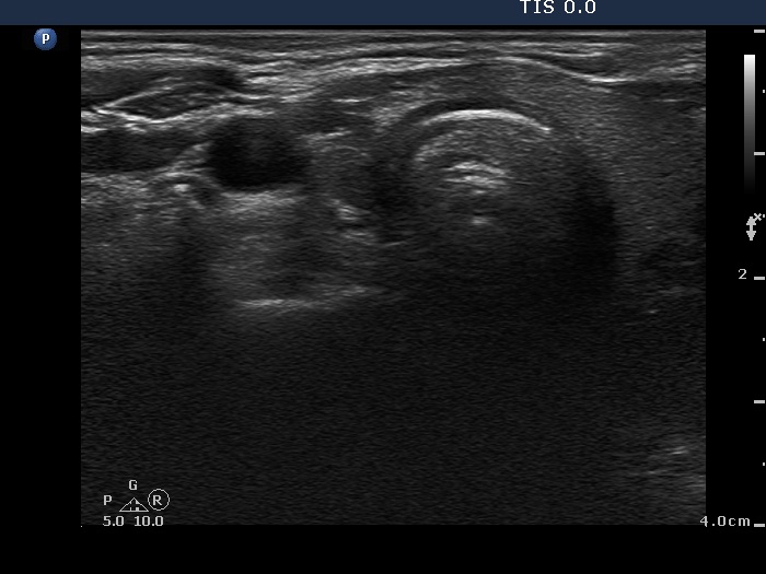
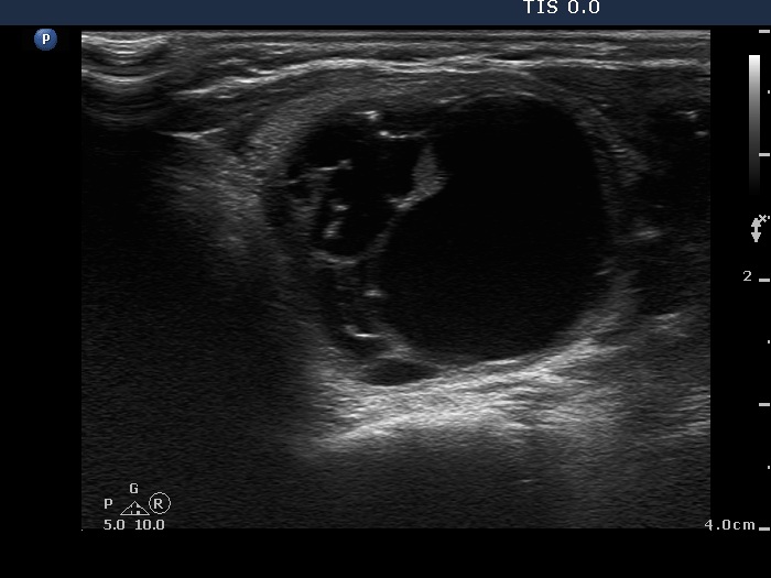
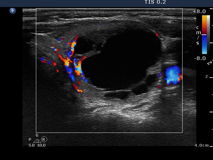
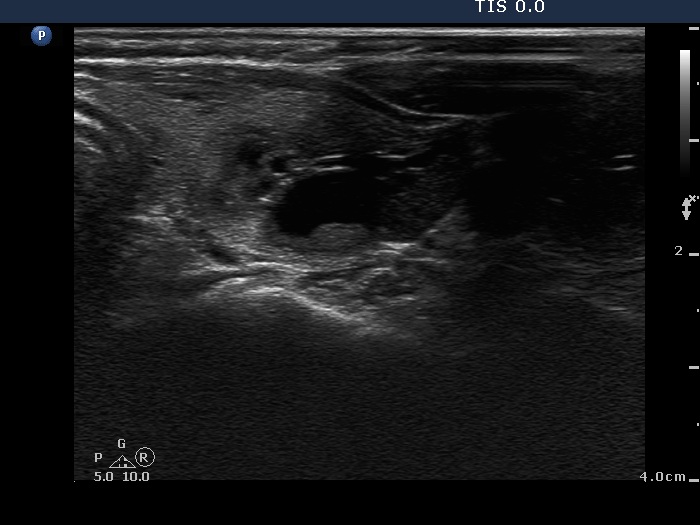
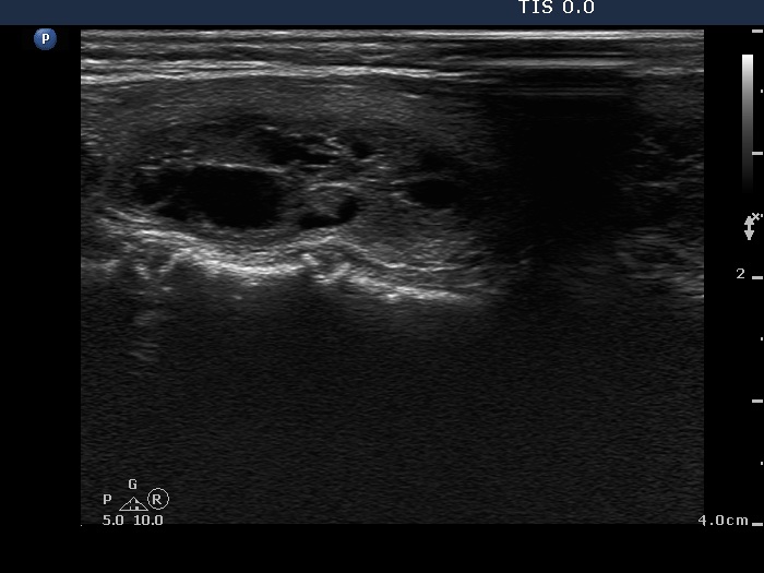
Ultrasonography. There was no parenchyma in the right the thyroid bed. The left lobe was minimally hypoechogenic and had a large, multichambered cystic nodule. There were both hyperechogenic lines and granules within the dorsal wall of the cystic areas. The solid part was moderately hypoechogenic.
8 mL serous fluid was removed, thereafter the solid part was aspirated. Cytology resulted in benign cystic lesion.
Comment. Most of the hyperechogenic figures correspond to posterior back wall enhancement. There were a few colloid crystals within the cystic fluid.
.