
Intranodular hyperechogenic figures - case 1197 (ultrasonographic picture 5)
|
|

|
|
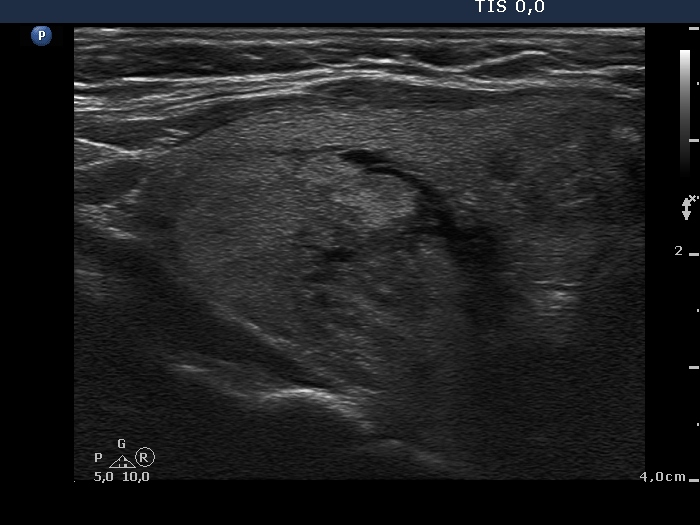
Left lobe, longitudinal scan. There is a minimally hypoechogenic, larger and a moderately hypoechogenic, smaller nodule, in the upper and in the middle part of the lobe, respectively.