
|
|
Intranodular hyperechogenic figures - case 1332
|
|


Clinical presentation: A 68-year-old man was referred for follow-up examination. She was first diagnosed having a nodular goiter more than 50 years ago. Occasionally she had "lump in the throat" feeling.
Palpation: a not firm nodule in the right lobe and a similar one in the isthmus.
Functional state: euthyroidism with TSH 0.61 mIU/L.
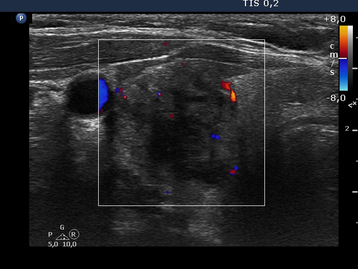
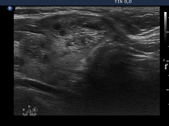
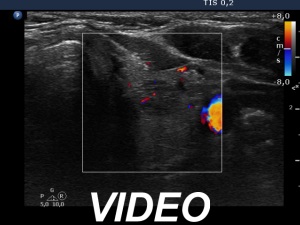
Ultrasonography. There were multiple nodules within the echonormal thyroid. The nodule in the right lobe had bright hyperechogenic granules while the nodules in the left lobe showed up synchronous echogenic lines and granules.
Aspiration cytology of the right lobe resulted in colloid goiter.
Comment. The bright echogenic foci in the right nodule are ambiguous figures, they might belong to punctate echogenic foci (microcalcifications).