
Intranodular hyperechogenic figures - case 1429 (ultrasonographic picture 4)
|
|

|
|
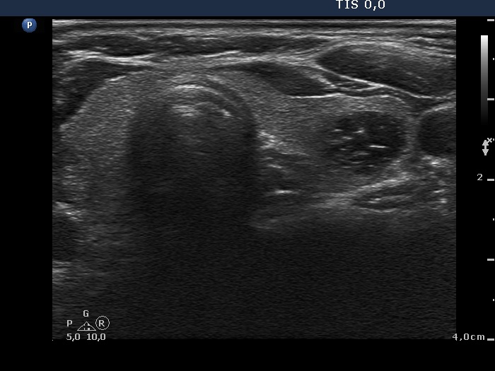
Left lobe, transverse view. There is a cystic lesion in the central part of the lobe. There are coexistent hyperechogenic lines and granules. These are either presentations of connective tissue or caused by posterior back wall enhancement.