
Intranodular hyperechogenic figures - case 1429 (ultrasonographic picture 5)
|
|

|
|
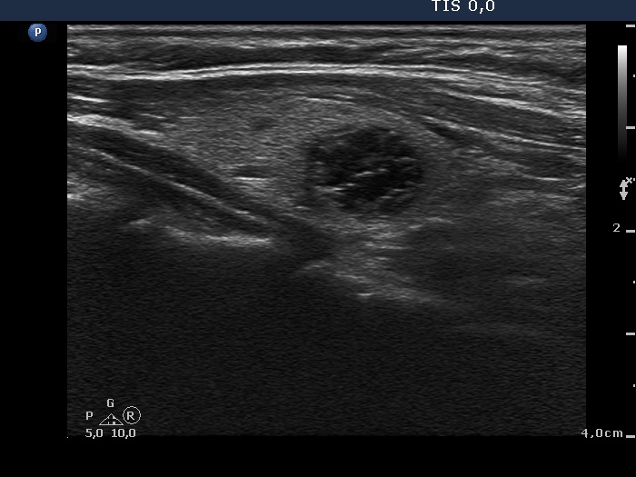
Left lobe, longitudinal view. This image proves that the lesion contains small cystic areas, therefore the intranodular figures belong to back wall cystic subgroup.