
|
|
Intranodular hyperechogenic figures - case 155
|
|

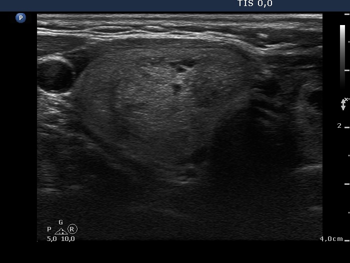


First examination (first row of images):
Clinical presentation: A 57-year-old woman was referred for aspiration cytology of a multinodular goiter has been known for more than two decades. The patient had no complaints.
Palpation: a multinodular goiter without any nodule suspicious on palpation.
Functional state: euthyroidism with TSH 0.96 mIU/L.
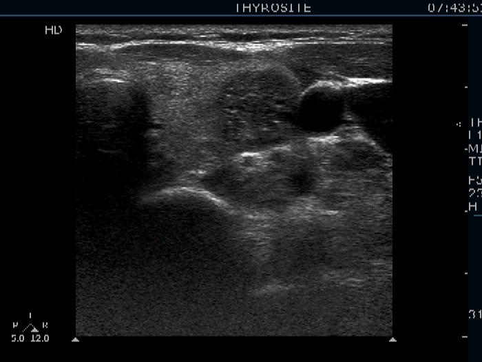
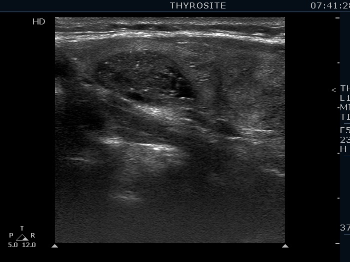
Ultrasonography. The thyroid was echonormal and was composed of multiple nodules. Most of them were hyperechogenic or minimally hypoechogenic. There was a hypoechogenic nodule presenting bright hyperechogenic granules in the upper part of the left lobe.
Cytology resulted in benign colloid goiter.
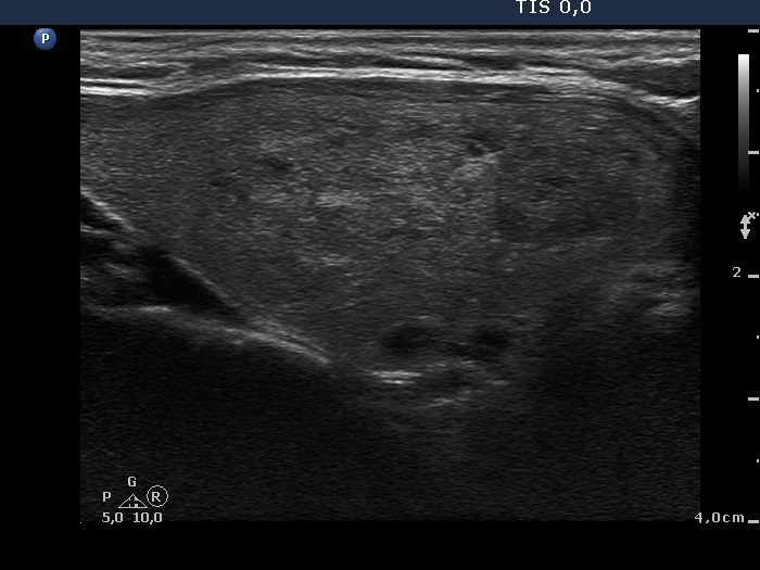
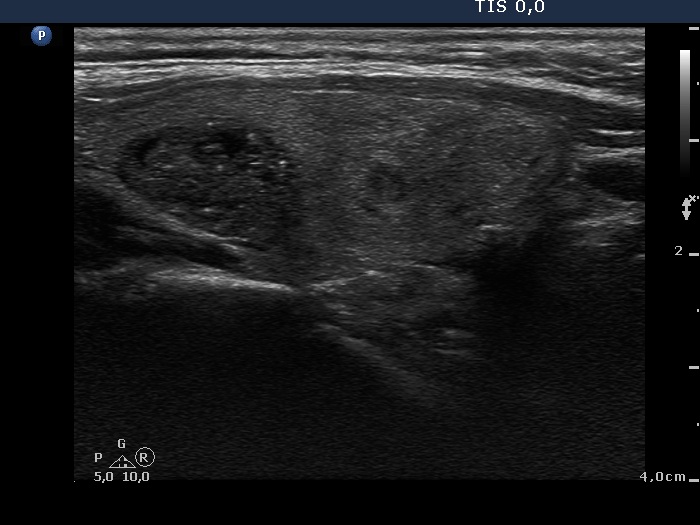
Second examination (second row of images):
Clinical presentation: The patient was referred for a follow-up examination.
Palpation remained unchanged.
Functional state: euthyroidism with TSH 2.11 mIU/L.
Ultrasonography. Both the size and the presentation of the nodules remained unchanged.
Comments.
-
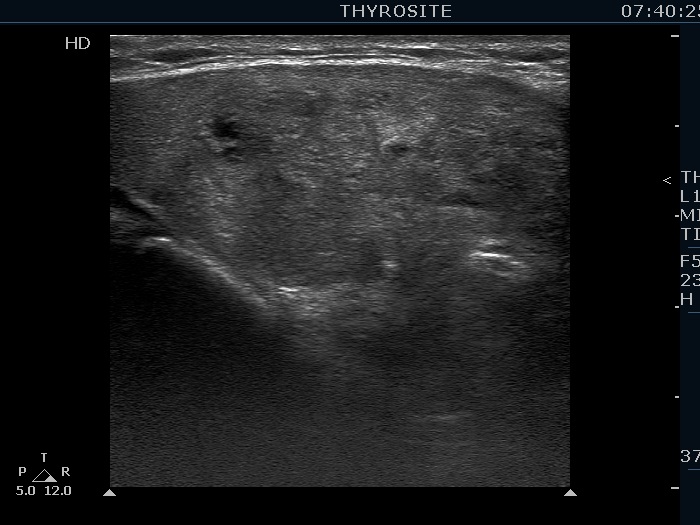
We may have doubt about the origin of echogenic figures, if we rely on the analysis of images. However, video proves that most of these granules and lines are related to tiny ventral cystic areas. It means that they are back wall cystic figures caused by posterior enhancement. Moreover, it is worth taking into account that the nodule lacks other suspicious characteristics which would be an unusual finding in the event of such large number of microcalcifications.
-
This study illustrates the difficulty of interpretation of hyperechogenic granules. In such cases the thorough analysis of the video is of much greater help than that of the images.