
|
|
Intranodular hyperechogenic figures - case 1644
|
|


Clinical presentation: A 42-year-old woman was referred for aspiration cytology of a thyroid nodule which was found on evaluation of thyroid enlargement.
Palpation: Both lobes were enlarged, no nodule could be palpated.
Functional state: euthyroidism with TSH 0.39 mIU/L, FT4 15.4 pM/L.
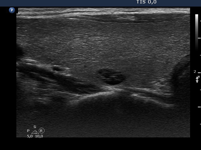
Ultrasonography. The thyroid was echonormal. There was a cystic lesion in the right while an inhomogeneous nodule in the left lobe. Both contained hyperechogenic figures.
Cytology of the nodule in the left lobe resulted in benign colloid goiter.
Comments. There is a striking similarity between back wall cystic figures and proliferation of connective tissue. Both present with synchronous presence of hyperechogenic lines and granules. There is only one difference between the two subgroups: the former occurs in the back wall of cystic lesions while the latter is independent of cystic fluid. On the other hand, the differentiation between these figures has a very limited importance. In this patient both types of echogenic figures can be observed.