
|
|
Intranodular hyperechogenic figures - case 1669
|
|


Clinical presentation: A 32-year-old woman was referred for aspiration cytology because a nodular goiter was diagnosed on evaluation of weight loss. Cytology was not diagnostic.
Palpation: an elastic nodule in the right lobe.
Results of blood tests: euthyroidism (TSH 1.71 mIU/L).
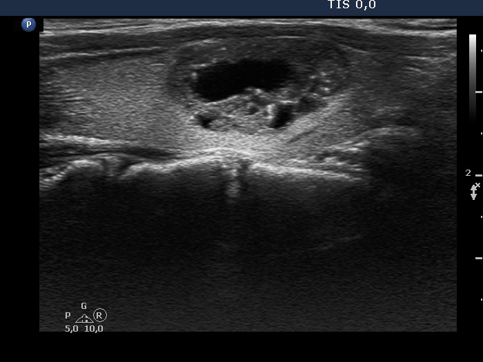
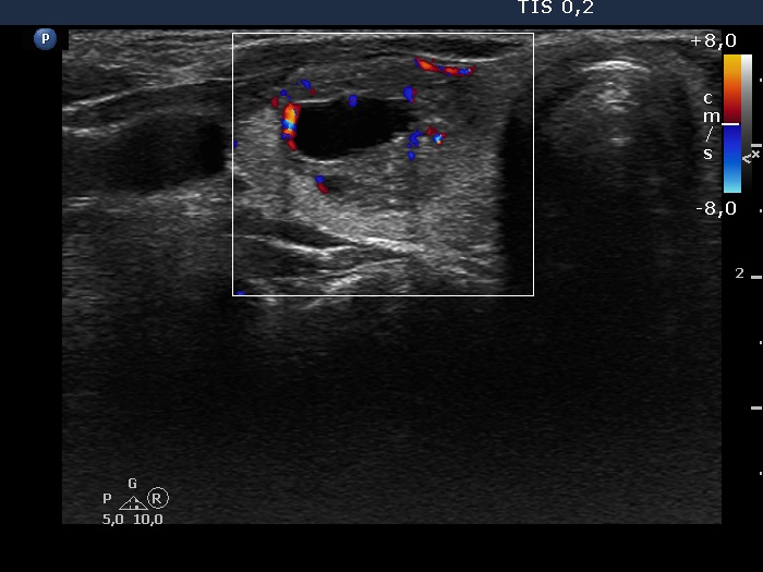
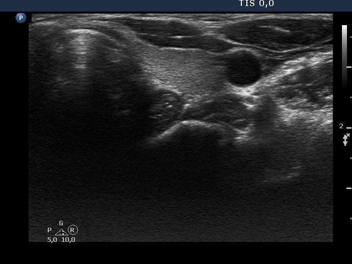
Ultrasonography. The thyroid was echonormal. There was a central-type cyst in the right lobe with partly hypoechogenic solid part having bright hyperechogenic granules. The lesion presented intranodular blood flow. There was another cystic lesion in the left lobe.
Cytology resulted in cystic degeneration.
Comment.
-
This case illustrates a not infrequent situation when we cannot differentiate comet-tail artifacts from microcalcifications.
-
On thorough analysis we can identify one granule which is clearly a comet-tail artifact and figures caused by posterior back wall enhancement.
-
The cyst in the right lobe was central-type which compared to a peripheral-type cyst, shares lower risk of malignancy.