
|
|
Intranodular hyperechogenic figures - case 638
|
|


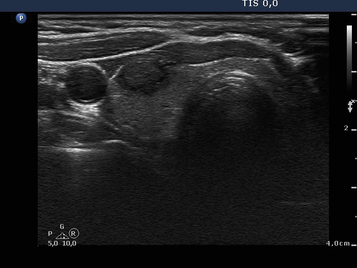
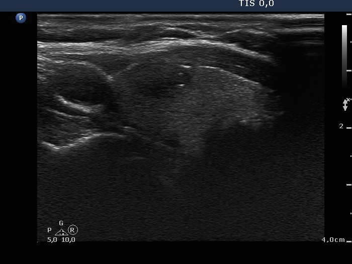


Clinical presentation: A 68-year-old woman was referred for an evaluation of a nodule discovered on ultrasonography. She was treated for breast cancer which gave metastasis to the lung.
Palpation: a small, hard nodule in the right lobe.
Functional state: euthyroidism with TSH 1.99 mIU/L.
Ultrasonography: There was a hypoechogenic nodule with cotton-like hyperechogenic patches and smaller punctate hyperechogenic granules in the right lobe. The nodule presented a type 3 vascular pattern, i.e. intranodular blood flow.
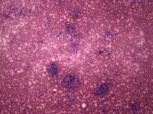
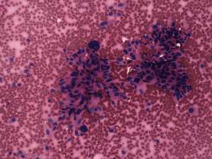
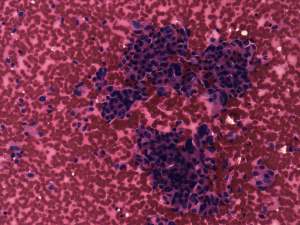
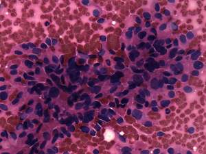
Cytological diagnosis: suspicion of medullary carcinoma.
Blood test for calcitonin: serum-level of calcitonin was 14.39 pM/L (normal value: 0-3.36).
Histopathology: medullary carcinoma.