
|
|
Intranodular hyperechogenic figures - case 401
|
|


Clinical presentation: A 76-year-old woman was referred for an evaluation of a newly discovered nodule. The patient noticed a lump in the left thyroid 4 months earlier. She had no complaints.
Palpation: a hard nodule in the left lobe.
Functional state: euthyroidism with TSH-level 1.40 mIU/L.
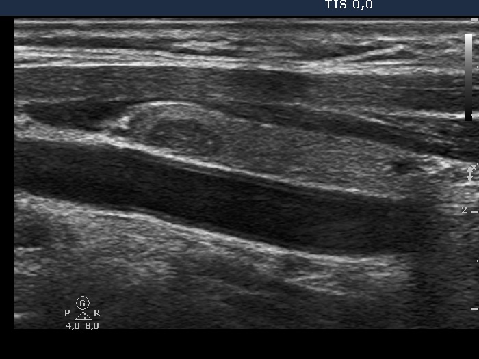
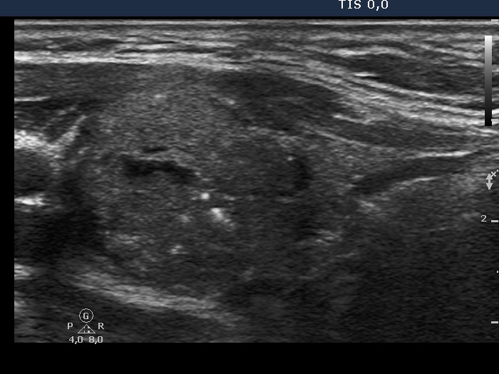
Ultrasonography. The thyroid was echonormal. There was a hypoechogenic nodule with microcalcifications in the right lobe, and another hypoechogenic nodule with microcalcifications and cotton-like patches in the left lobe.
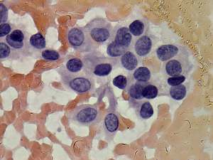
Cytological diagnosis.
Second row of images: papillary cancer in the right nodule.
Third row of images: medullary cancer in the left nodule.
Blood test for calcitonin: serum-level of calcitonin was 5.91 pM/L (normal value: 0-3.36).
Histopathology: papillary cancer in the right and medullary cancer in the left lobe.
Comments.
-
This is a very edifying case as regards the presentation of hyperechogenic figures. Considering the histopathology, these in the right nodule are very likely microcalcifications. On the other hand, they are closer to a non-specific granule.
-
There are larger patch-like hyperechogenic figures in the left, medullary carcinoma focus. These are clearly amyloid deposits. While the relatively smaller and more bright figures are larger than a microcalcification. They are probably also amyloid deposits.