
Intranodular hyperechogenic figures - case 808
Follow-up investigation 5 years after the first visit (ultrasonographic picture 9)
|
|

|
|
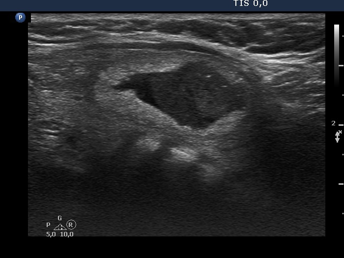
Left side of the isthmus, longitudinal scan - after aspirating 5 mL serous fluid. There are several bright hyperechogenic lines and granules. These might be presentations of connective tissue or back wall figures caused by posterior enhancement.