
Intranodular hyperechogenic figures - case 808
Follow-up investigation 5 years after the first visit (ultrasonographic picture 10)
|
|

|
|
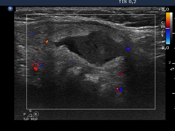
Left side of the isthmus, transverse scan, color Doppler mode - after aspirating 5 mL serous fluid. The vascularization is scanty.