
|
|
Intranodular hyperechogenic figures - case 928
|
|


Clinical presentation: An 85-year-old woman was referred for evaluation of a nodular goiter has been known for decades. The patient had already difficulties in swallowing for several years. CT scan disclosed substernal spread of the right thyroid. The trachea was narrowed to half. There were multiple nodules with normal uptake on scintigraphy.
Palpation: a multinodular goiter in the right lobe.
Functional state: euthyroidism with TSH 0.31 mIU/L, FT4 15.9 pm/l, FT3 4,63 pM/L.
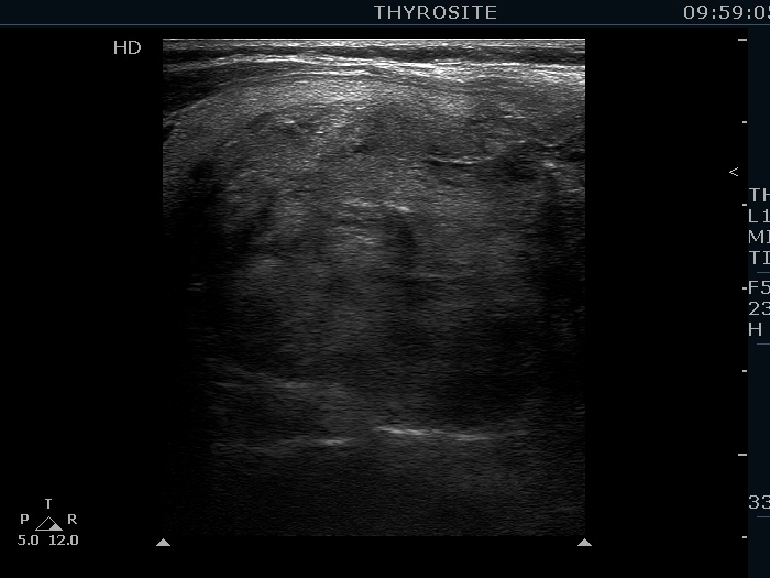
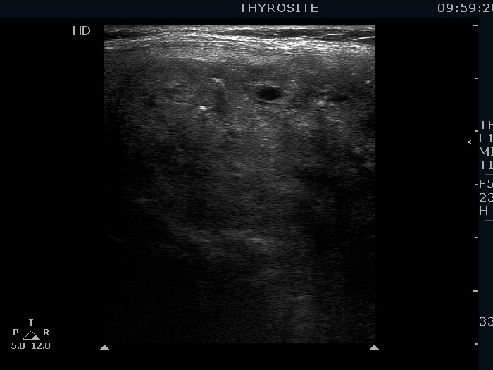
Ultrasonography. The thyroid was echonormal. There were several nodules in the right lobe including a large echonormal which presented coarse calcification, synchronous echogenic lines and granules and a few brighter punctate echogenic foci, as well.
Aspiration cytology resulted in benign colloid goiter.
A right lobectomy was performed. Histopathology disclosed benign hyperplastic nodules.