
|
|
Intranodular hyperechogenic figures - case conp 052
|
|


Clinical data: A 66-year-old woman was referred for aspiration cytology because of a nodule detected on PET-CT scan. She was operated on ductal invasive breast carcinoma for a year.
Palpation: a firm nodule in the left lobe.
Functional state: euthyroidism (TSH 0.49 mIU/L).
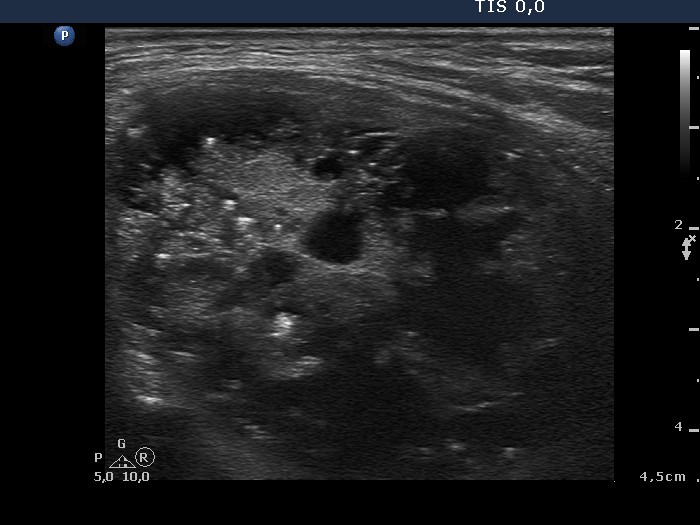
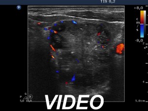
Ultrasonography. The thyroid was echonormal. A large mixed nodule occupied almost the entire left lobe. The nodule presented numerous microcalcifications, taller-than-wide sign and a star-like central solid area. Although the solid part of the lesion was echonormal and the vascularization was not specific, the presentation of the nodule was very suspicious for malignancy.
Three aspirations were performed. In all cases a minimal amount of brownish fluid was gained. There were only macrophages on the cytological smear.
Wash out thyroglobulin level exceeded 478 µg/L.
Our final combined clinical-ultrasound-cytological was papillary carcinoma with great probability.
Histopathology disclosed papillary carcinoma.
Comment.
-
The ultrasound presentation itself is highly suspicious for a papillary carcinoma.
-
Video record proved that the nodule had back wall cystic figures, microcalcifications and some of the hyperechogenic figures could not be interpreted other than comet-tail artifact.