
|
|
The echogenicity of the nodule - case 2152
|
|

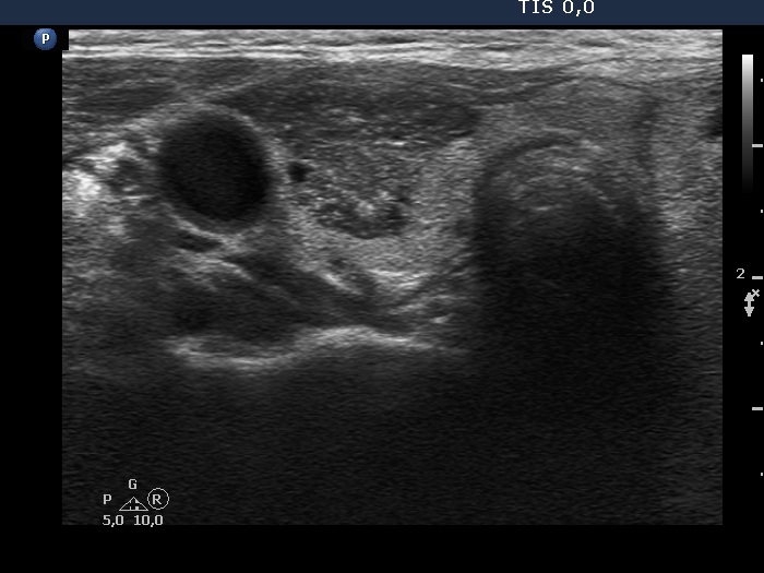


Clinical presentation: An 83-yr-old woman was referred for evaluation of multinodular goiter which has been known for decades.
Palpation: Both lobes were nodular, none of the nodules was firm.
Laboratory tests: TSH 0.89 mIU/l, FT4 13,5 pM/L.
Ultrasound. The right lobe had a dominantly hypoechoic, heterogeneous nodule. There was an undulation on the medial border which was caused by an echonormal lesion. The left lobe presented a large dominantly iso/hyerechoic nodule showing cystic areas.
Aspiration cytology from the hypoechoic nodule resulted in benign colloid goiter.
Comments.
- A nodule should follow the anatomy of the thyroid, which frequently narrows near to the carotid artery. This situation was responsible of a non-pathological spiculation demonstrated in this case.
- It is evident, that an impression on the nodule border caused by another lesion does not correspond to pathological lobulation.