
Halo sign and vascular pattern of nodules - case 2248 (ultrasonographic picture 3)
|
|

|
|
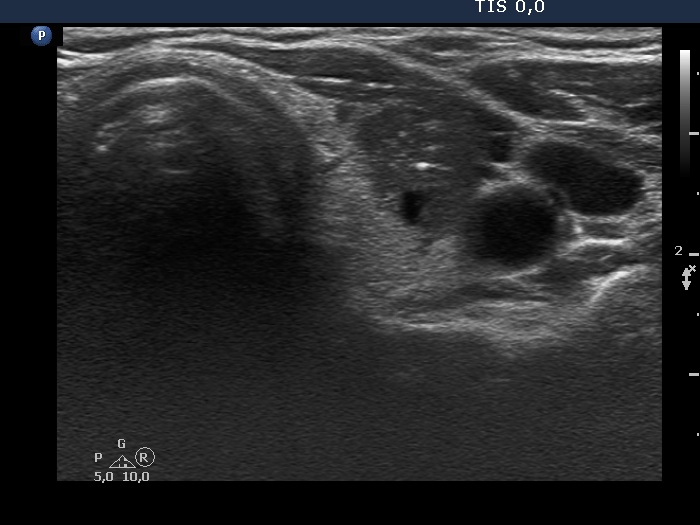
Left lobe, transverse view. There is a deeply hypoechoic nodule with irregular borders. The nodule shows bulging and abutting contours. The bright echogenic granule cannot be classified other than a microcalcification.