
Halo sign and vascular pattern of nodules - case 2248 (ultrasonographic picture 4)
|
|

|
|
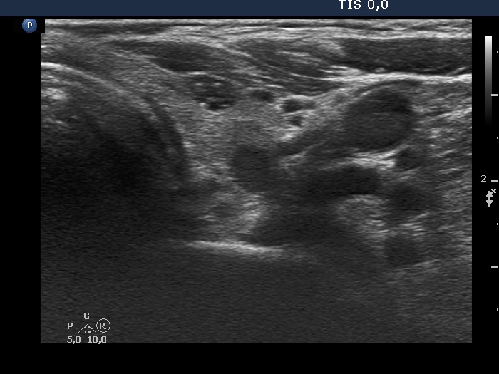
Lower part of the left lobe, transverse scan. There are two more hypoechic lesions. The dorsal one shows taller-than-wide shape. The echogenic figures in the ventral lesion are related to ventral cystic areas, therefore these are back wall figures.