
|
|
Halo sign and vascular pattern of nodules - case 2254
|
|

Clinical data: A 29-year-old woman requested an evaluation because of difficulties in swallowing.
Palpation: a hard nodule in the upper part of the right lobe.
Laboratory test: TSH 1.47 mIU/L.
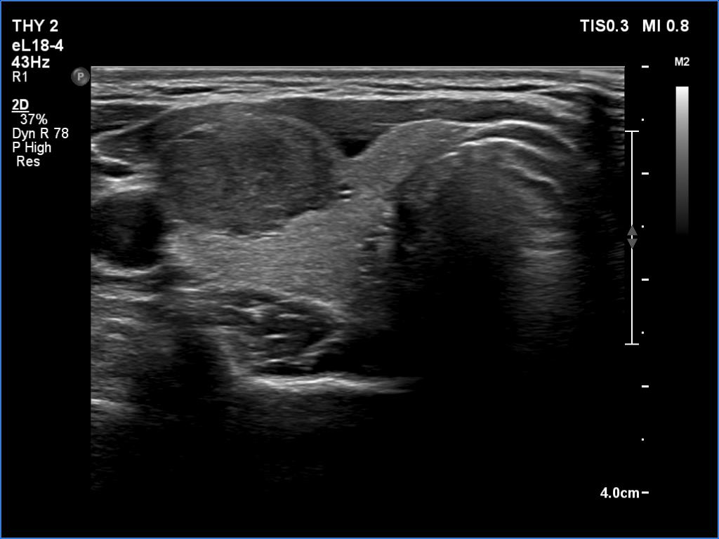
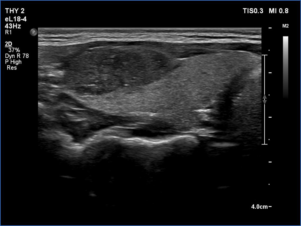
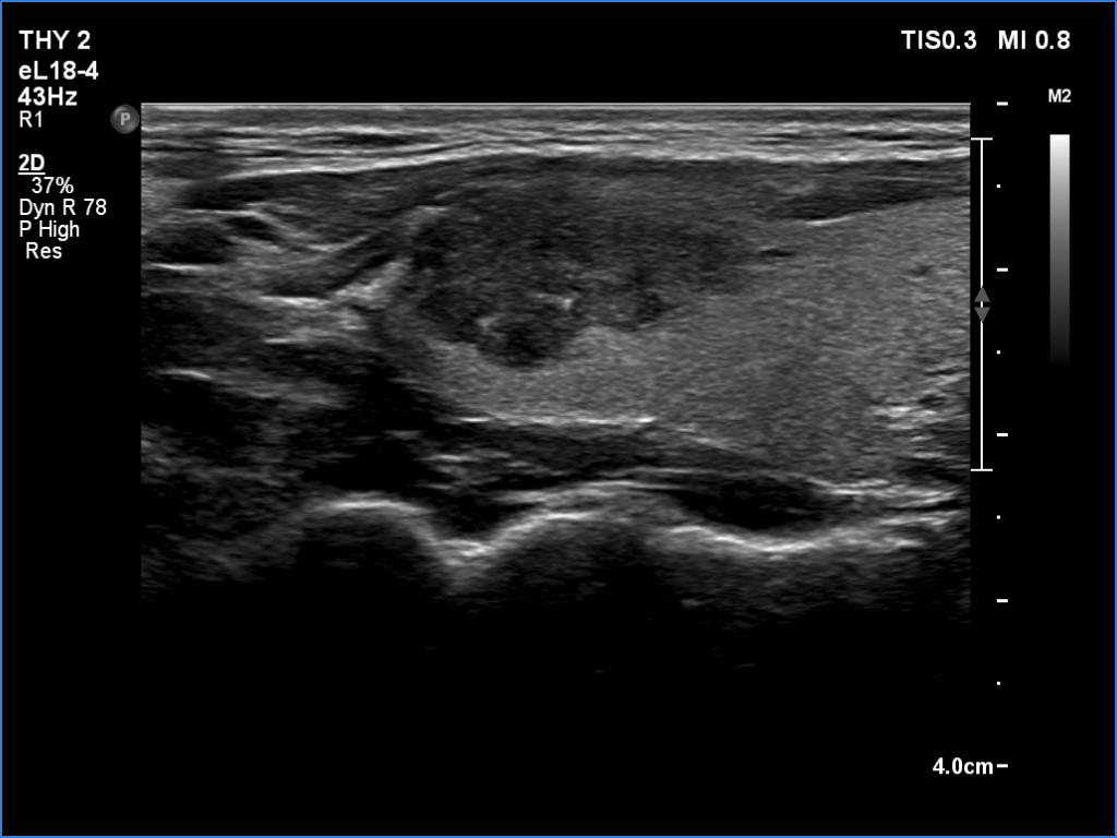
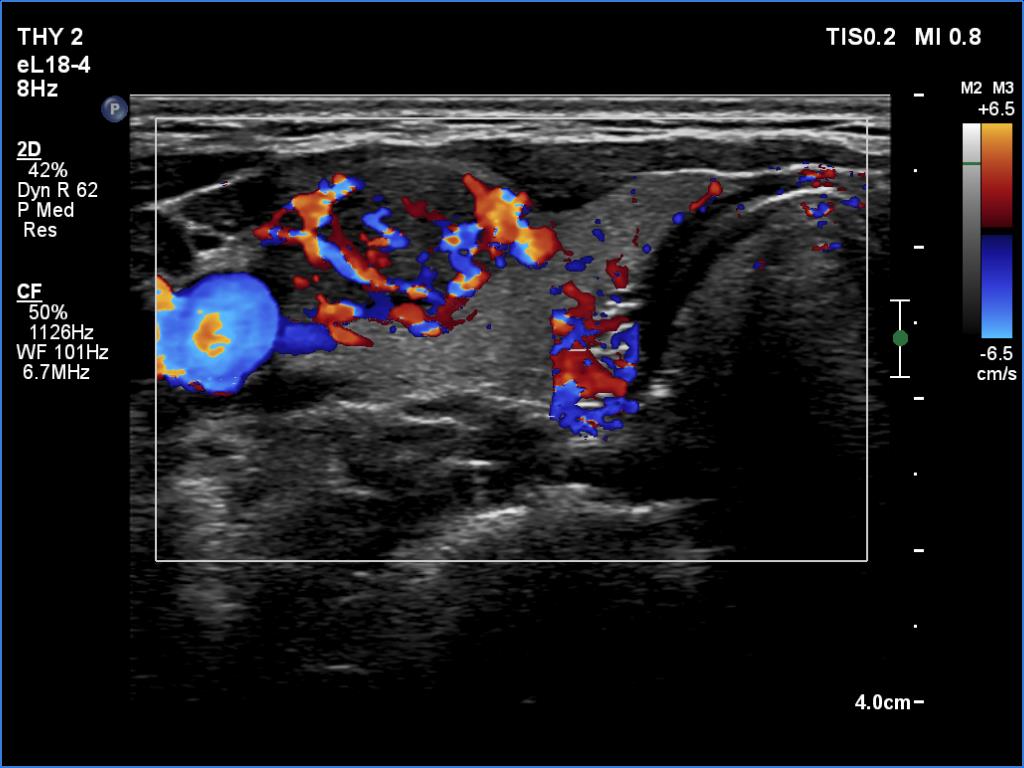
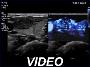
Ultrasonography. The thyroid was echonormal and presented two discrete lesions in the right lobe. The larger, upper one showed irregular margins, abutting and bulging contours and had back wall figures. The intranodular vascularization was significantly increased. The smaller lesion had microcalcifications and presented taller-than-wide shape.
Cytology of the larger nodule resulted in papillary carcinoma.
Total thyroidectomy was performed. Histopathology disclosed a T1b papillary cancer.
Comment. It is worth thoroughly analyzing the intranodular echogenic figures. These are partly linear and most of them are related to tiny ventral cystic areas.