
|
|
Halo sign and vascular pattern of nodules - case 444
|
|


Clinical data: A 41-year-old woman was sent for aspiration cytology. She discovered a lump in the right thyroid 3 months before the examination.
Palpation: a firm nodule in the right lobe.
Hormonal evaluation: euthyroidism with TSH 1.86 mIU/L.
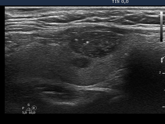
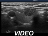
Ultrasonography revealed a hypoechogenic nodule which predominantly had echogenic granules but in a smaller number echogenic lines, as well. These were found mostly dorsal to tiny cystic areas, therefore these very likely corresponded to back wall cystic figures. Nevertheless, the presence of microcalcification cannot be fully excluded. The nodule did not present halo sign and had intranodular blood flow.
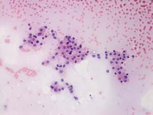
Cytology itself was very suspicious for a Hürthle-cell tumor.
A combined ultrasound-cytological diagnosis was benign Hürthle-cell metaplasia with less than 1% risk of carcinoma.
We advised regular follow-up instead of surgery. The patient decided to undergo on surgery.
Histopathology disclosed benign hyperplastic nodules with extensive oxyphilic metaplasia.
Comment.
This is an excellent example for the potential of a new way of thinking. Both the ultrasound and the cytological pattern was itself suspicious but not of the same tumor. The increased intranodular blood flow and the possibility of microcalcifications could raise the possibility of a papillary carcinoma. However, it was very unlikely on cytological pattern because of the lack of inclusions and grooves. And conversely, the cytological pattern corresponded to a follicular-type oxyphilic tumor, however considering the lack of halo sign and perinodular blood, the risk of a follicular tumor was surely less than 5%.