
Halo sign and vascular pattern of nodules - case conp 068
Follow-up investigation seven years later (ultrasonographic picture 4)
|
|

|
|
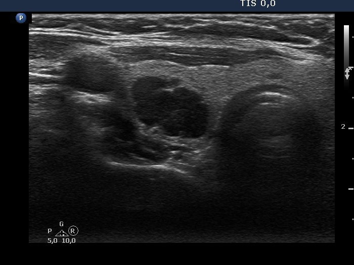
Middle part of the right lobe, transverse scan. There is a deeply hypoechoic lesion which proved to be a parathyroid adenoma on histopathology.